
Aneurysm Clipping
Brain aneurysm clipping is a type of microsurgery in which a titanium surgical clip is used to close off an aneurysm in the brain. The surgeon makes a small opening in your skull to reach your brain. They use an operating microscope and very small instruments to perform detailed surgical procedures. Precision is key during microsurgery to avoid harming nerves, blood vessels and brain tissue. Clipping may be performed on a ruptured or unruptured aneurysm.
The Procedure
Aneurysm clipping (craniotomy) is performed under general anesthesia in an operating room. The neurosurgeon:
- Stabilizes the head so it cannot move during surgery.
- Makes an incision in the scalp and the skin is pulled out of the way.
- Opens a small window of bone (bone flap) in the skull through which the brain can be accessed.
- Opens the dura, the protective covering around the brain.
- Isolates the blood vessel and aneurysm from the surrounding brain tissue.
- Places a metal clip across the aneurysm’s neck. More than one clip may be used.
- Stitches the dura closed.
- Reattaches the bone flap to the skull using thin metal plates and screws and returns the skin flap over it.
- Closes the scalp incision with stitches.


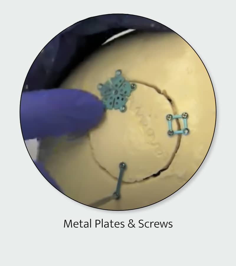
The clip effectively cuts off blood flow from the artery to the aneurysm and remains in place permanently, as do the metal plates and screws (under the scalp) used to reattach the bone flap.
Patients who had an unruptured aneurysm typically stayed in the hospital for two days.
Patients who had a ruptured brain aneurysm will stay in the hospital for two to three weeks. During that time, patients are monitored for serious side effects possible after a bleed in the brain. These include vasospasm, hydrocephalus, problems with the heart, lungs, or other conditions.
Most aneurysm clips these days are MR-compatible, meaning they can safely enter an MRI machine. However, you should double-check with your surgeon to make sure. The plates and screws that close the craniotomy bone flap site are MR-compatible.
Disclaimer
The Maine Brain Aneurysm Awareness Committee (MBAAC) does not provide medical advice and does not recommend, endorse or make any representation about the efficacy, appropriateness or suitability of any specific tests, products, procedures, treatments, services, opinions, health care providers or other information that may be contained on or available through this website or our social media.
NEVER DISREGARD PROFESSIONAL MEDICAL ADVICE OR DELAY SEEKING MEDICAL TREATMENT BECAUSE OF SOMETHING YOU HAVE READ ON OR ACCESSED THROUGH THIS WEBSITE.
MBAAC’s website and social media contains information that is intended to educate patients and their caregivers about brain aneurysms. It is not intended to be a substitute for professional medical advice, diagnosis or treatment. It is crucial that care and treatment decisions related to vascular malformations of the brain and any other medical condition be made in consultation with a doctor or other qualified medical professional. Articles that MBAAC utilize are not vetted by any medical professionals associated with MBAAC. We use our discretion to choose topics that may be of interest to members of our community.