Endovascular Coiling/Stent
Neurosurgeons can now perform minimally invasive endovascular approaches to treat some brain aneurysms. Endovascular coiling and stent embolization use specialized technology to treat an aneurysm from inside the blood vessel.
This procedure is typically performed in the radiology suite by either a neurosurgeon or an interventional radiologist.
Generally, a coiling and stent procedure follows this process:
- You will be positioned on your back on the X-ray table.
- An intravenous (IV) line will be started in your hand or arm.
- You will be connected to an electrocardiogram (ECG) monitor that records the electrical activity of the heart. Your vital signs (heart rate, blood pressure, and breathing rate) and neurological signs will be monitored during the procedure.
- A catheter may be inserted into your bladder to drain urine.
- The radiologist or neurosurgeon will check your pulses below the groin site where the catheter will be inserted and mark them with a marker so that the circulation to the limb below the site can be checked after the procedure.
- The skin over the injection site will be cleansed. A local anesthetic will be injected.
- A small incision will be made in the skin to expose the artery in the groin or neck.
- A catheter will be inserted into the artery in your groin or neck using a guide wire. The catheter will be guided through the blood vessel into the brain using fluoroscopy (a special type of X-ray, similar to an X-ray “movie”).
- Once the catheter has been guided to the affected artery in the brain, contrast dye will be injected to make the aneurysm and surrounding blood vessels visible on X-ray.
- The aneurysm will be measured and its shape and other characteristics will be recorded.
- Next, a smaller catheter will be inserted into the initial catheter.
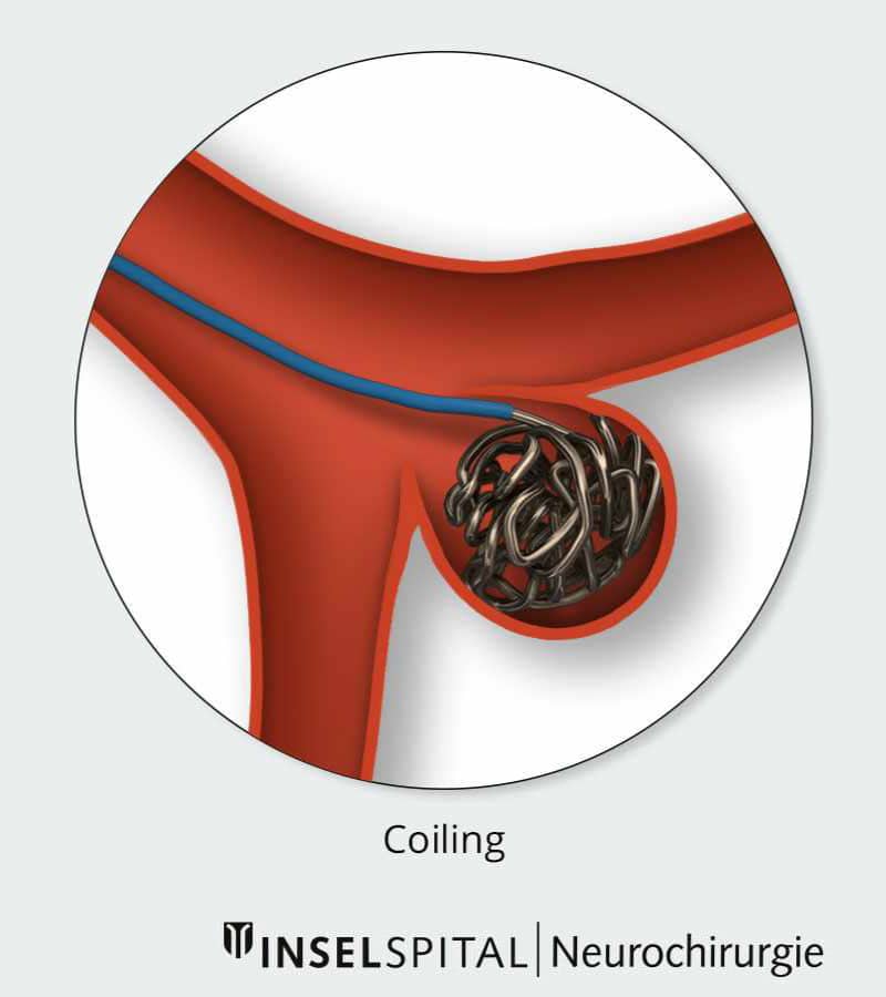
- Once the catheter has reached the aneurysm, the healthcare provider will manipulate the coil into the aneurysm.
- When the coil has been completely inserted into the aneurysm, the coil is separated from the catheter.
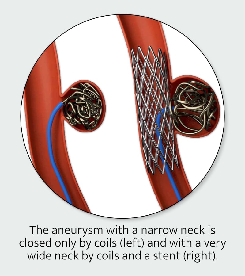
- The healthcare provider will insert as many coils as needed to completely seal off the aneurysm. The coils will form a mesh-like structure inside the aneurysm.
- After the aneurysm has been “packed” with coils, additional X-ray images will be taken to make sure the aneurysm has been sealed off. The coil is left in place permanently in the aneurysm.
- Once the aneurysm has been sealed off, the catheter will be removed. After the insertion site stops bleeding, a dressing will be applied.

First two Images: University Clinic for Neurosurgery, Inselspital Bern © CC BY-NC 4.0
Disclaimer
The Maine Brain Aneurysm Awareness Committee (MBAAC) does not provide medical advice and does not recommend, endorse or make any representation about the efficacy, appropriateness or suitability of any specific tests, products, procedures, treatments, services, opinions, health care providers or other information that may be contained on or available through this website or our social media.
NEVER DISREGARD PROFESSIONAL MEDICAL ADVICE OR DELAY SEEKING MEDICAL TREATMENT BECAUSE OF SOMETHING YOU HAVE READ ON OR ACCESSED THROUGH THIS WEBSITE.
MBAAC’s website and social media contains information that is intended to educate patients and their caregivers about brain aneurysms. It is not intended to be a substitute for professional medical advice, diagnosis or treatment. It is crucial that care and treatment decisions related to vascular malformations of the brain and any other medical condition be made in consultation with a doctor or other qualified medical professional. Articles that MBAAC utilize are not vetted by any medical professionals associated with MBAAC. We use our discretion to choose topics that may be of interest to members of our community.